
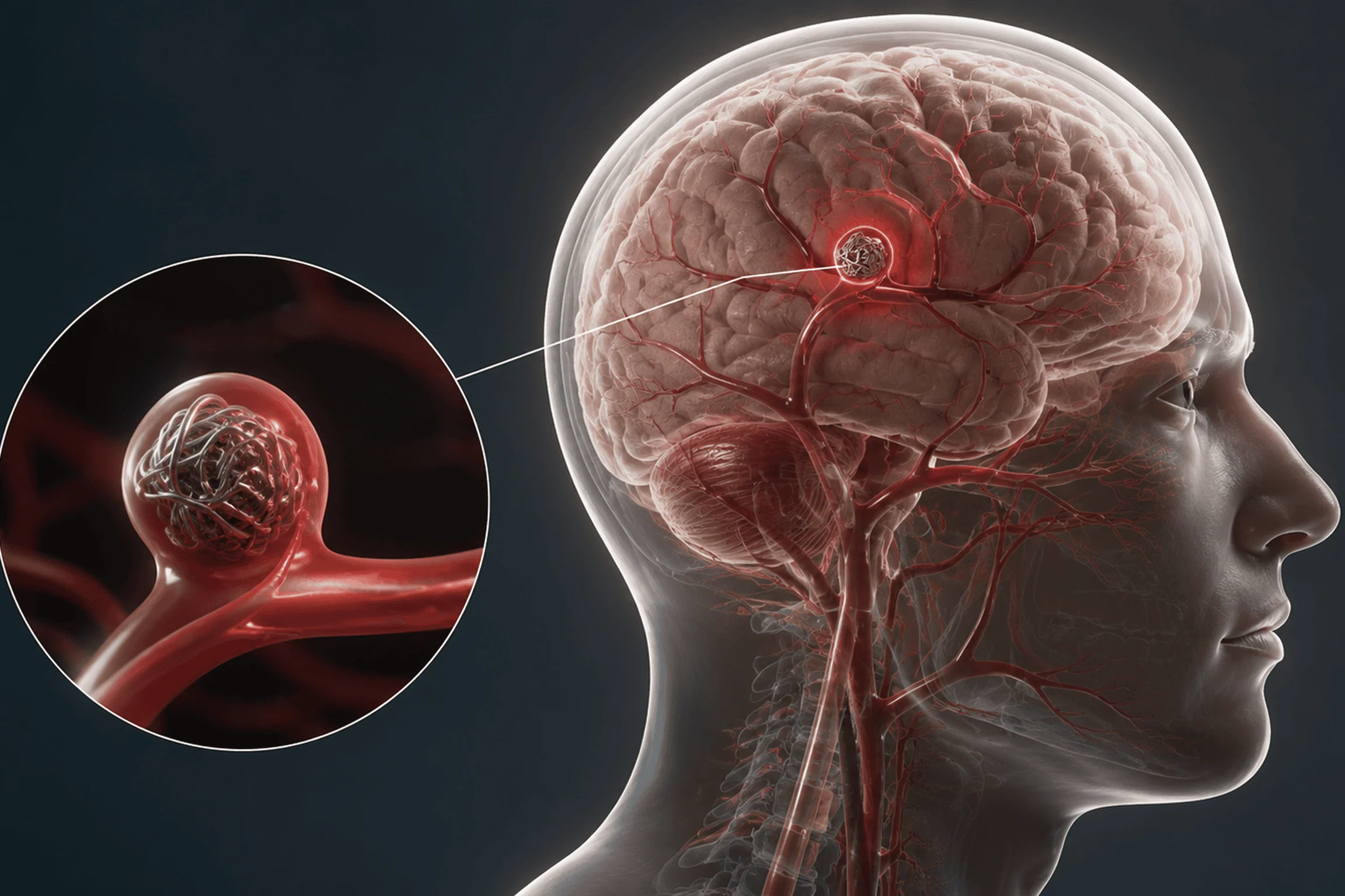
Brain Aneurysm Management
- 24×7 Neuro-Emergency Response
- Advanced Cerebral Angiography
- Endovascular Coiling & Flow-Diversion Centre
- Expert Neuro-Interventionalists
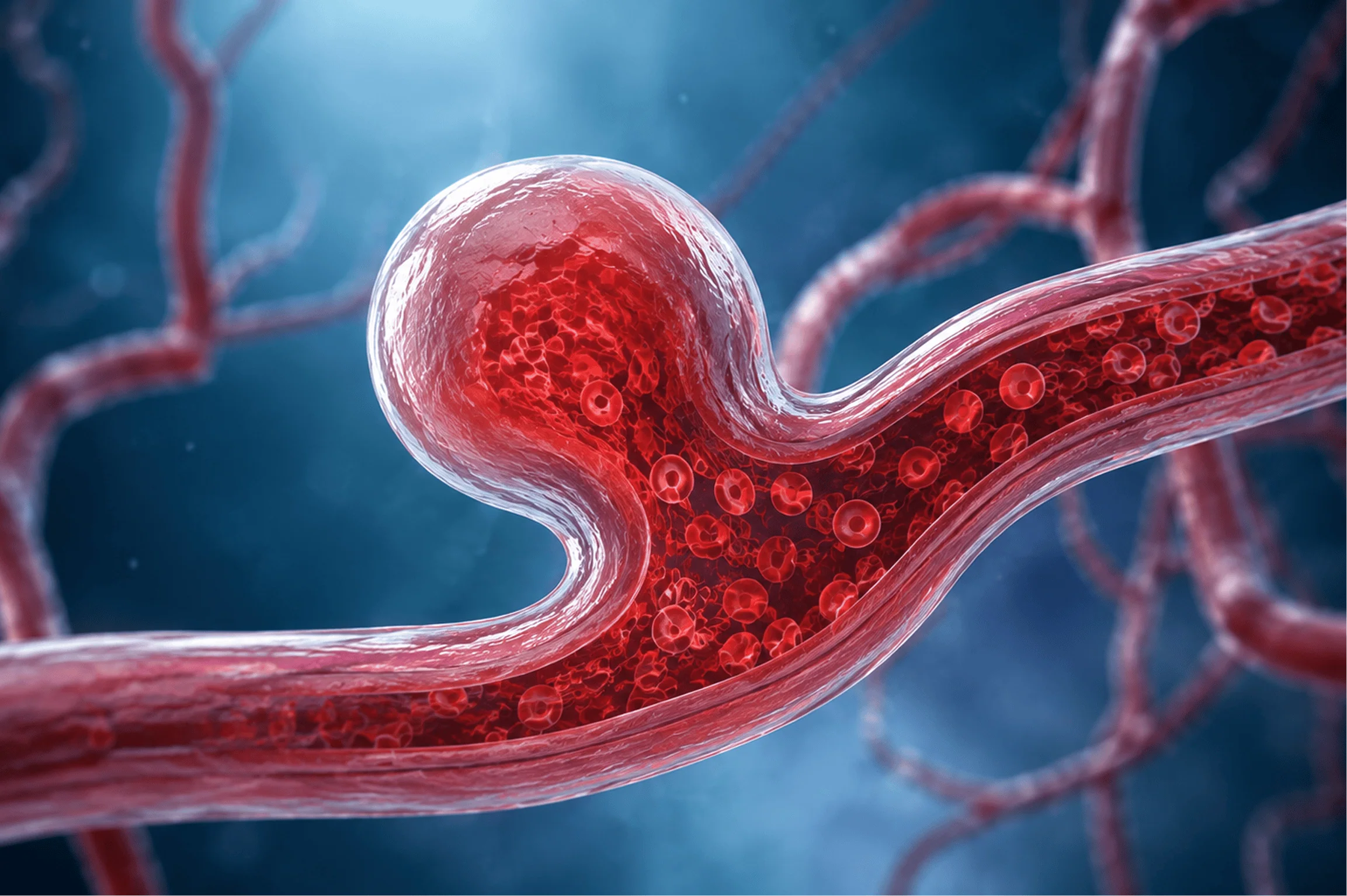
What is a Brain Aneurysm?
A brain aneurysm forms when the wall of a cerebral artery weakens, allowing blood pressure to push outward and create a localised bulge. Many aneurysms remain silent for years causing no symptoms until they enlarge or rupture. This is precisely why early imaging and careful clinical monitoring are essential. When identified, many aneurysms can be secured through minimally invasive endovascular techniques, significantly reducing the risk of rupture and its potentially catastrophic consequences.
Silent risk
Rupture danger
Early detection
Treatable condition
Symptoms & Warning Signs
- Sudden, severe headache often described as the worst headache of one's life.
- Blurred or double vision.
- Nausea and vomiting.
- Sensitivity to light.
- Drooping eyelid, dilated pupil, or facial numbness.
- Seizures or loss of consciousness, particularly in the setting of rupture.
Remember the Warning Signs
S
Sudden Severe Headache
A headache of unprecedented severity — often described as a thunderclap — is a cardinal sign of aneurysm rupture. Treat it as an emergency.
V
Vision Changes
Sudden blurring, double vision, or a drooping eyelid may indicate pressure from an enlarging aneurysm on surrounding neural structures.
N
Neck Stiffness
Stiffness or pain in the neck following a severe headache may signal bleeding around the brain and warrants immediate investigation.
T
Time to Call
If any of these signs are present, seek emergency care without delay. Early intervention is the single most important factor in outcome.
The Treatment Journey
Faster Diagnosis
Minimally Invasive Treatment
Structured Neuro-Emergency Response
Integrated Care
Post‑Procedure Care & Recovery
Following aneurysm treatment, care shifts immediately to neurological stabilization, structured rehabilitation, and long-term surveillance.
Immediate Recovery
Post-procedure monitoring in ICU or high-dependency unit, with continuous neurological observation to detect vasospasm, re-bleeding, or early complications.
Home Care Plan
A tailored rehabilitation plan addressing neurological recovery, activity, and medications for gradual return to daily function.
Follow-Up Consultation
Scheduled MR angiography or digital subtraction angiography to confirm durable aneurysm occlusion and guide long-term management.

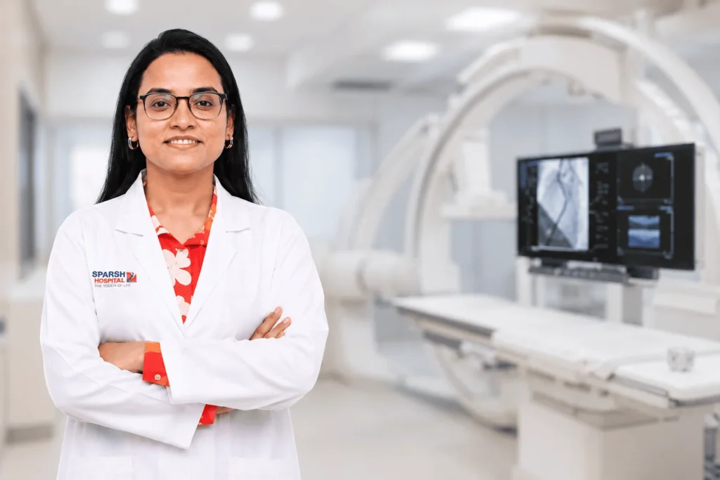
Why Choose Dr. Deepmala Karmakar?
Our interventional radiology practice brings specialised expertise across the full spectrum of vascular and non-vascular care. Every case begins with a thorough clinical evaluation ensuring the most appropriate, evidence-based intervention is selected for each patient’s specific condition.
Every procedure is performed under real-time advanced imaging whether ultrasound, fluoroscopy, or CT enabling precise access and targeted treatment with minimal disruption to surrounding tissue and faster recovery outcomes.
Experienced Specialists
Each procedure is led by a trained interventional radiologist with deep expertise across vascular and non-vascular domains.
Advanced Technology
Procedures are guided by state-of-the-art imaging infrastructure, ensuring accuracy at every step of the intervention.
24×7 Response
Time-critical conditions including acute stroke and vascular emergencies are managed with round-the-clock readiness and a dedicated response protocol.
Image-Guided Precision
Targeted access under real-time imaging minimises tissue disruption, reduces procedure time, and supports faster, more predictable recovery.
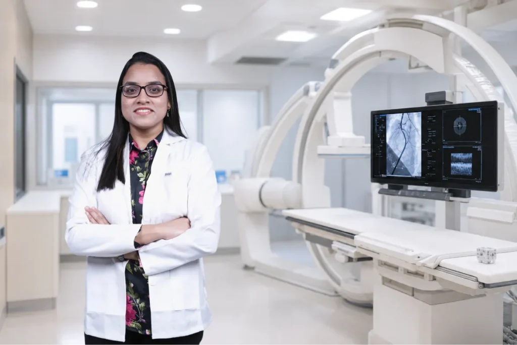
Why Choose
Dr. Deepmala Karmakar?
Brain aneurysm management demands a precise combination of advanced imaging capability, cerebrovascular expertise, and the technical proficiency to execute endovascular procedures safely in complex anatomical settings. Our neuro-interventional practice is equipped to assess, stratify, and treat aneurysms across the full spectrum of presentations unruptured, symptomatic, and ruptured.
Experienced Specialists
Procedures are led by a trained neuro-interventionalist with dedicated expertise in cerebrovascular anatomy, endovascular coiling, and flow-diversion techniques.
Advanced Imaging Technology
High-resolution cerebral angiography and neuro-imaging infrastructure support accurate aneurysm characterisation and precise, image-guided intervention.
Neuro-Emergency Pathways
Structured emergency protocols enable rapid assessment and treatment for suspected aneurysm rupture, with streamlined triage-to-angiography workflows.
Image-Guided Precision
Real-time fluoroscopic and angiographic guidance ensures accurate device deployment within the aneurysm, supporting complete and durable occlusion.
Frequently Asked Questions
What causes a brain aneurysm?
Brain aneurysms develop from a combination of factors including chronic hypertension, smoking, a family history of aneurysms, and inherent structural weakness in the arterial wall which may in some cases be present from birth. Certain connective tissue disorders and polycystic kidney disease are also associated with a higher incidence.
What is endovascular coiling?
Endovascular coiling is a minimally invasive procedure performed under angiographic guidance, in which a catheter is navigated through the arterial system to the site of the aneurysm. Tiny platinum coils are then deployed within the aneurysm sac to promote clotting, eliminate blood flow into the aneurysm, and reduce the risk of rupture without the need for open cranial surgery in most cases.
Can an unruptured aneurysm be safely monitored instead of treated?
Yes, in carefully selected cases. Small aneurysms with favourable characteristics low-risk morphology, stable size, and absence of symptoms may be managed conservatively with periodic imaging surveillance rather than immediate intervention. The decision is made through an individualised risk assessment that weighs the aneurysm's specific features against the patient's age, health status, and overall risk profile.
Get in Touch
Every Second Counts
98%
Successful Procedures
15 min
Door-to-Imaging Time
24x7
Emergency Response
10+
Years of Experience
