
Aortic Interventions
- Rapid Aortic Emergency Response
- Advanced CT & Angiographic Imaging
- Endovascular Stent-Graft Centre
- Expert Vascular Interventional Team
What are Aortic Interventions?
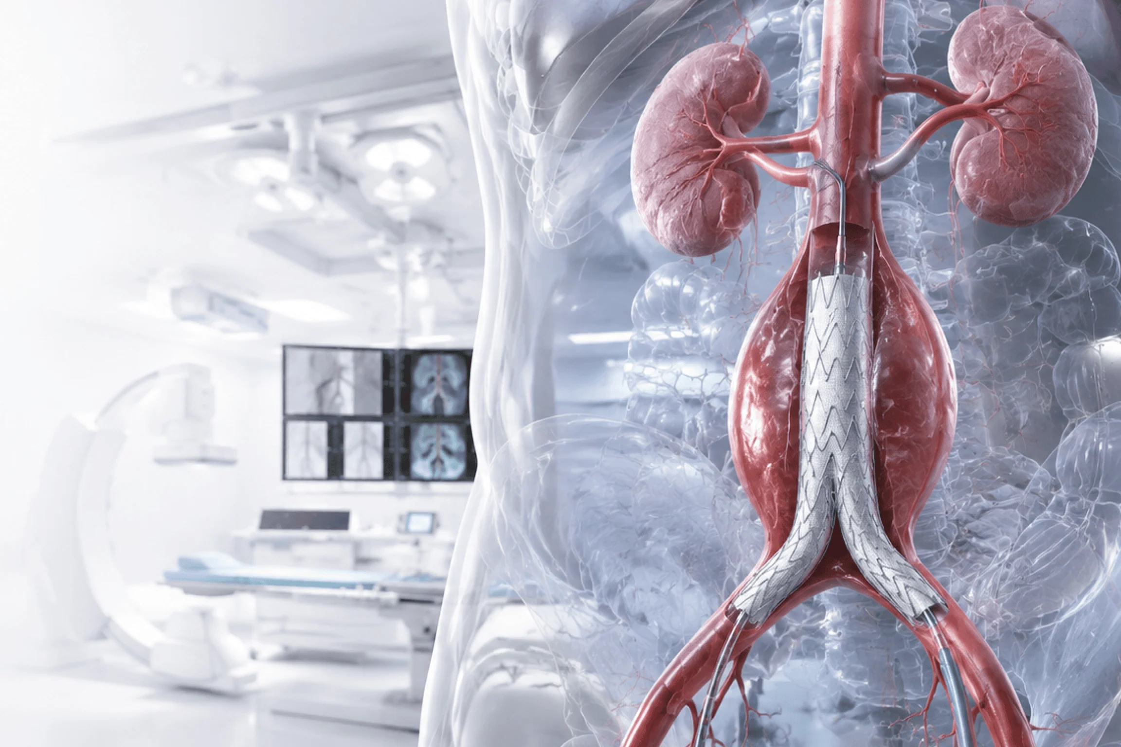
The aorta carries blood from the heart to the rest of the body. When its wall weakens, bulges, or tears — through aneurysm, dissection, or occlusive disease — prompt specialist evaluation is essential. Endovascular repair reinforces the damaged segment from within using a catheter-delivered stent-graft, avoiding open surgery in most suitable cases.
Silent risk
Rupture danger
Early detection
Endovascular Repair
Symptoms & Warning Signs
- Sudden, severe chest, back, or abdominal pain — often described as tearing or ripping in character.
- A pulsating sensation or noticeable throbbing mass in the abdomen.
- Sudden breathlessness, dizziness, or loss of consciousness.
- Unequal pulses or blood pressure readings between the arms.
- Persistent chest, back, or abdominal pain that does not resolve.
Conditions Treated
- Abdominal Aortic Aneurysm (AAA) — aneurysmal dilatation of the aorta below the diaphragm.
- Thoracic Aortic Aneurysm (TAA) — aneurysmal disease involving the thoracic aorta.
- Aortic Dissection — a tear in the inner layer of the aortic wall allowing blood to track between layers.
- Aortic Occlusive Disease — blockages within the aorta or its branches impairing distal blood flow.
Remember the Warning Signs
S
Sudden Severe Pain
Chest, back, or abdominal pain of sudden onset — particularly tearing or ripping in character — is a cardinal sign of aortic emergency. Act immediately.
P
Pulsating Abdomen
A noticeable throbbing mass in the abdomen may indicate an enlarging aortic aneurysm requiring urgent evaluation.
W
Sudden Weakness
Fainting, acute breathlessness, or unequal pulses between the arms may signal aortic dissection or rupture.
T
Time to Call
If any of these signs are present, seek emergency care without delay. In aortic emergencies, speed of intervention is directly linked to survival.
Aortic Treatment Journey
Faster Diagnosis
Precise Anatomical Planning
Minimally Invasive Repair
Integrated Follow-Up Care
Post‑Procedure Care & Recovery
Immediate Recovery
Post-procedure care is delivered in an ICU or high-dependency unit, with continuous haemodynamic and neurological monitoring to detect and manage early complications including endoleak or access site issues.
Home Care Plan
Detailed discharge guidance is provided covering physical activity restrictions, blood pressure management, and medication review — tailored to the specific procedure performed and the patient's vascular risk profile.
Follow-Up Consultation
Scheduled CT or ultrasound imaging is arranged at defined intervals to confirm stent-graft stability, assess for endoleak, and guide long-term aortic surveillance in line with established vascular guidelines.

Why Choose Dr. Deepmala Karmakar?
Our interventional radiology practice brings specialised expertise across the full spectrum of vascular and non-vascular care. Every case begins with a thorough clinical evaluation ensuring the most appropriate, evidence-based intervention is selected for each patient’s specific condition.
Every procedure is performed under real-time advanced imaging whether ultrasound, fluoroscopy, or CT enabling precise access and targeted treatment with minimal disruption to surrounding tissue and faster recovery outcomes.
Experienced Specialists
Each procedure is led by a trained interventional radiologist with deep expertise across vascular and non-vascular domains.
Advanced Technology
Procedures are guided by state-of-the-art imaging infrastructure, ensuring accuracy at every step of the intervention.
24×7 Response
Time-critical conditions including acute stroke and vascular emergencies are managed with round-the-clock readiness and a dedicated response protocol.
Image-Guided Precision
Targeted access under real-time imaging minimises tissue disruption, reduces procedure time, and supports faster, more predictable recovery.
Why Choose
Dr. Deepmala Karmakar?
Every case is planned using high-resolution CT aortography, with meticulous anatomical assessment to ensure appropriate device selection and durable long-term outcomes.
Experienced Specialists
Procedures are led by a trained vascular interventionalist with dedicated expertise in endovascular aortic anatomy, stent-graft selection, and complex aortic repair techniques.
Advanced Imaging Technology
High-resolution CT, MR, and angiographic imaging infrastructure supports precise anatomical planning and accurate intraoperative device deployment.
Aortic Emergency Pathways
Structured emergency protocols enable rapid clinical assessment, imaging, and treatment planning for patients presenting with suspected aortic rupture or dissection.
Image-Guided Precision
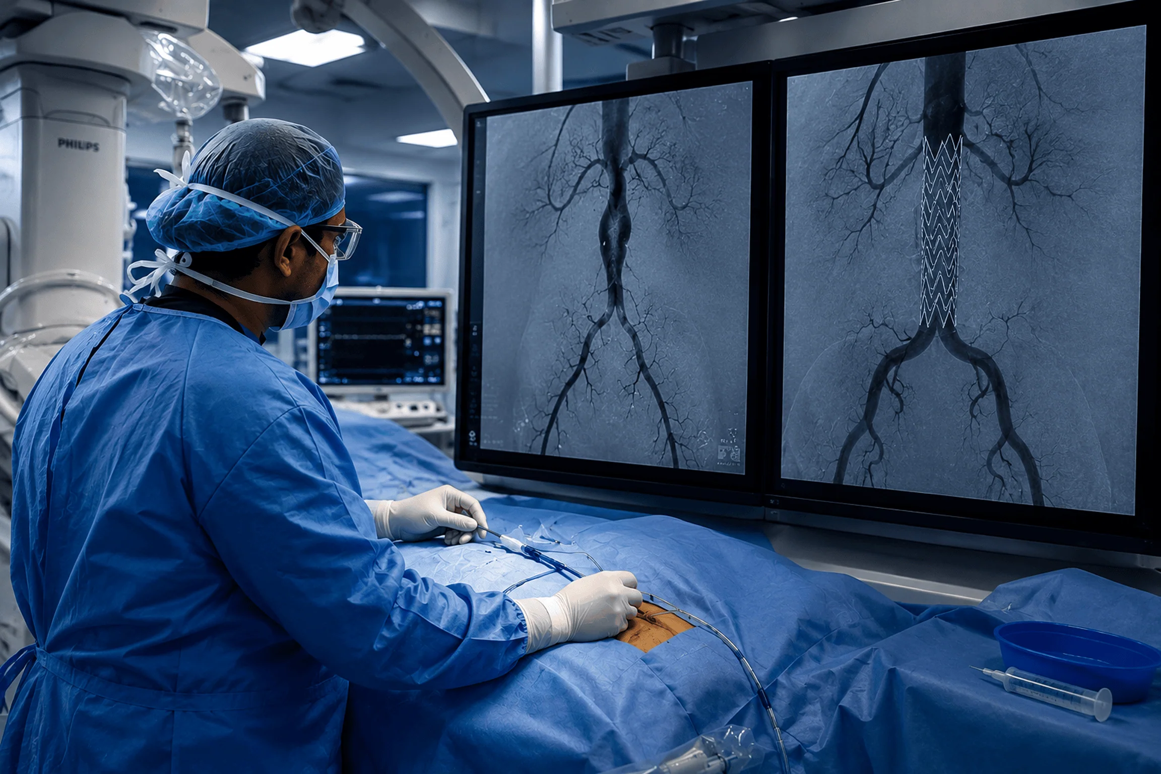
Real-time fluoroscopic and angiographic guidance throughout stent-graft deployment ensures accurate positioning, complete aneurysm exclusion, and immediate assessment of technical outcome.
Frequently Asked Questions
What is an aortic aneurysm?
An aortic aneurysm is a localised, abnormal dilatation of the aortic wall — occurring when the structural integrity of the vessel wall is compromised and blood pressure causes it to bulge outward. Aneurysms may enlarge progressively over time and carry a risk of rupture if not appropriately monitored and treated.
What is endovascular stent-graft repair?
Endovascular stent-graft repair is a minimally invasive procedure in which a fabric-covered metal stent is delivered via catheter through the femoral arteries and deployed precisely within the aorta to reinforce the weakened or diseased segment from within. It excludes the aneurysm or dissection from arterial blood flow without the need for open chest or abdominal surgery in most suitable cases.
Can an aortic aneurysm be monitored instead of treated immediately?
Yes, in carefully selected cases. Small aneurysms below established size thresholds with stable growth rates are managed conservatively with periodic imaging surveillance and risk factor modification — including blood pressure control and smoking cessation. Larger, rapidly enlarging, or symptomatic aneurysms warrant timely intervention.
Get in Touch
When the Aorta Is at Risk, Precision Is Everything
98%
Successful Procedures
15 min
Door-to-Imaging Time
24x7
Emergency Response
10+
Years of Experience
